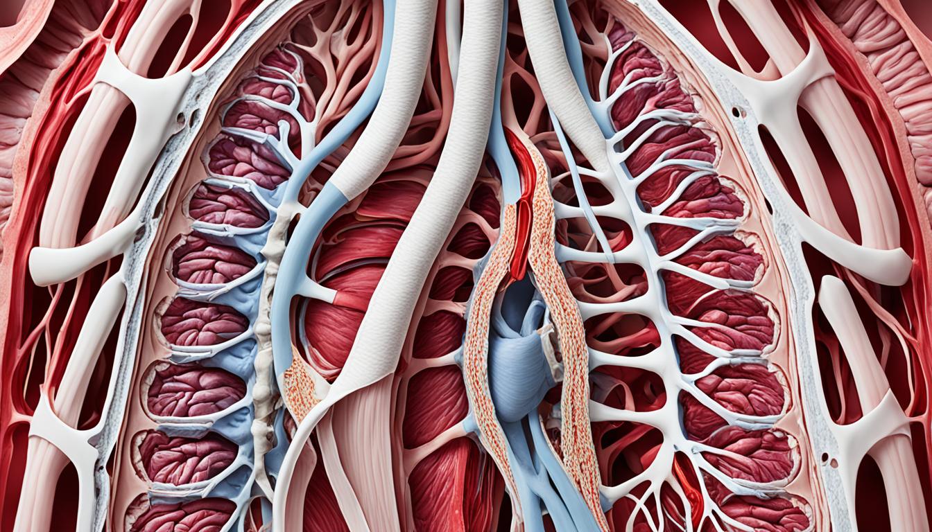
Thoracic aortic aneurysm is a rare type that mainly affects the chest area. It can be linked to certain diseases that affect the body’s connective tissues like Marfan and Ehlers-Danlos syndromes. Reasons for it can be a build-up of plaque in the arteries, a severe blow, or inflammation. Things like high blood pressure, smoking, or inherited risks can make someone more likely to develop this issue. Doctors usually find it through scans such as CT or MRI. They might fix it with an operation or try new treatments like stem cells.
Key Takeaways:
- Thoracic aortic aneurysm is a less common type of aortic aneurysm that primarily affects the thorax.
- Connective tissue disorders such as Marfan and Ehlers-Danlos syndromes, or congenital bicuspid aortic valve are often associated with thoracic aortic aneurysm.
- Atherosclerosis, blunt injury, and inflammation are the main causes of thoracic aortic aneurysm.
- Risk factors for thoracic aortic aneurysm include hypertension, smoking, and genetic factors.
- Diagnosis involves imaging tests such as CT scans or MRI.
- Treatment options for thoracic aortic aneurysm may include surgery or stem cell therapy.
Causes and Risk Factors of Thoracic Aortic Aneurysm
Thoracic aortic aneurysm is a significant health concern. It can come from many factors such as health problems and lifestyle. Knowing these causes helps find issues early and avoid them.
A common cause is atherosclerosis. This is when plaque builds up in arteries. The plaque can weaken the aorta’s walls, making an aneurysm more likely.
Accidents and trauma can also be a cause. They can damage the aortic wall. This makes aneurysms more likely to happen.
Inflammation is key in aortic arch aneurysm development. It can harm the aorta’s connective tissue. This weakens the walls, upping the risk for an aneurysm.
People with certain disorders are at higher risk. Conditions like Marfan or Ehlers-Danlos weaken artery walls. This makes aneurysms more likely.
Bad habits like smoking and high blood pressure are also risky. They stress the arteries. This stress can lead to aneurysms.
A small number of cases are from family genes. About 10% have a family history of aortic aneurysms. Keeping an eye on genetic risks is important.
But not everyone with these risks will get an aneurysm. Regular check-ups and symptom awareness are key. They help spot and treat issues early.
Diagnosis and Treatment of Thoracic Aortic Aneurysm
To diagnose a Thoracic Aortic Aneurysm (TAA), doctors use imaging tests. They look at the aneurysm’s size and where it is. CT scans and MRI are the most common tests. They help doctors know the aneurysm’s size and severity.
Early diagnosis is key for better treatment outcomes. The size, location, and patient’s health guide the treatment. Surgery is the usual step to fix the aorta.
Diagnostic Imaging Techniques
CT scans show detailed images of the chest area. They tell us about the aneurysm’s size and shape. This information is important for planning the right treatment.
MRI creates detailed aorta images without radiation. It also looks at the aneurysm closely.
Treatment Options
Surgery fixes big, dangerous aneurysms. Doctors repair with synthetic grafts. This surgery stops the aneurysm from getting bigger.
Stem cell therapy is new and uses the body’s own cells. It aims to fix the aorta without surgery. This therapy is a major hope for patients.
Both surgery and stem cell therapy aim to better patients’ lives with this condition.
Diagnostic Imaging Techniques for Thoracic Aortic Aneurysm
| Imaging Technique | Advantages | Disadvantages |
|---|---|---|
| CT scan (Computed Tomography) | – Detailed images of the aorta – Shows aneurysm size and place well – Quick to finish |
– Uses radiation – Some patients react to contrast dye – Doesn’t show all aorta wall parts well |
| MRI (Magnetic Resonance Imaging) | – Safe and detailed look at the aorta – No radiation used |
– Takes more time – Not good for some patients with metal implants – Small space can be uncomfortable for some |
The table above lists benefits and downsides of CT scans and MRIs for aneurysm diagnosis. It shows what these tests are good at and not so good at. This helps doctors pick the best tests for each case.
Using these advanced tests helps doctors pick the right treatment. Whether it’s surgery or new stem cell therapy, early detection and treatment are crucial for better patient outcomes.
Conclusion
Thoracic aortic aneurysm is a severe health issue needing quick diagnosis and fitting treatment. The key to spotting this issue early is using high-tech imaging like CT scans and MRIs. These methods show the aneurysm clearly, helping doctors to plan treatment.
There are many ways to treat this condition. One common method is surgery to fix or replace the damaged part of the aorta. But now, stem cell therapy is becoming a popular option. It aims to repair the tissue, potentially offering a less invasive way to treat the problem.
Fast treatment is key because the aneurysm can tear or burst, which is very dangerous. Luckily, with the advancements in medical technology, the outlook for these patients is getting better. Now, more than ever, there is hope for a brighter future and improved quality of life for those with thoracic aortic aneurysms.

