Pulmonary atresia with intact ventricular septum (PA-IVS) is a rare heart condition affecting less than 1% of heart patients. It is caused by a blockage in the heart’s right ventricle. Usually, this blockage might be either a small hole (membranous) or a missing path (muscular). The exact reason for PA-IVS is not fully known, but it happens during the early stages of a baby’s growth.
About 4 to 8 babies out of 100,000 are born with PA-IVS. Thanks to new ways to check babies before they’re born, like fetal echocardiography, doctors can now spot PA-IVS early and more accurately. They use echocardiography, a special heart ultrasound, as the main test to find PA-IVS. If needed, doctors might also do a cardiac catheterization to learn more.
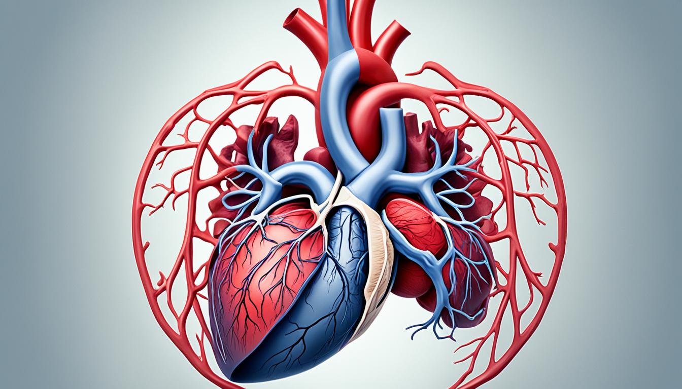
Learning about PA-IVS symptoms, causes, and treatments is key to helping those with the condition. In this article, we will find out more about what PA-IVS looks like inside the heart. We’ll also see how doctors check for it and the ways they can treat it, like using stem cell therapy.
Key Takeaways:
- PA-IVS is a rare congenital heart defect, making up less than 1% of heart issues.
- The blockage in PA-IVS can be a small hole or a missing path in the heart’s right ventricle.
- Doctors often find PA-IVS using echocardiography. They might do a cardiac catheterization to learn more.
- PA-IVS can be treated with surgery, procedures to help the heart work better, or stem cell therapy.
- It’s very important for people with PA-IVS to visit their doctors regularly for care.
Characteristics and Anatomy of Pulmonary Atresia with Intact Ventricular Septum
Pulmonary atresia with intact ventricular septum (PA-IVS) has unique characteristics. These traits are key to diagnosing and treating it.
RVOT Obstruction: In PA-IVS, there’s a blockage in the right ventricle’s outflow tract. This block can be at the valve or muscle level. It stops blood from flowing from the right ventricle to the lungs, affecting how well the lungs get blood.
RV Morphology: The right ventricle in PA-IVS can have different shapes. Sometimes it looks like it has three parts. But it might be just two or one part if some areas are too big or blocked.
Tricuspid Valve Abnormalities: People with PA-IVS often have issues with their tricuspid valve. This valve can be too small or not formed right. It might not open or close properly. These issues affect blood flow from the heart’s upper chamber to the lower one.
Coronary Artery Abnormalities: The higher pressure in the heart’s right ventricle can cause problems in the coronary arteries for PA-IVS patients. These problems might affect how well blood flows to the heart muscle. In some cases, blood flow might even depend on it flowing backward from the right ventricle.
Anatomy of Pulmonary Atresia with Intact Ventricular Septum
| RVOT Obstruction | Obstruction in the right ventricular outflow tract |
|---|---|
| RV Morphology | Variations in the structure of the right ventricle |
| Tricuspid Valve Abnormalities | Small and dysplastic tricuspid valve with stenosis and/or regurgitation |
| Coronary Artery Abnormalities | Abnormal connections and stenoses within the coronary arteries |
Grasping the details of PA-IVS is vital for its care. It helps doctors make plans that are right for each patient’s needs.
Clinical Presentation and Evaluation of Pulmonary Atresia with Intact Ventricular Septum
Understanding the signs and symptoms of pulmonary atresia with intact ventricular septum (PA-IVS) is key. It’s important for getting the right diagnosis and treatment. This part looks at how doctors spot and evaluate PA-IVS.
Presentation and Symptoms
PA-IVS often shows through cyanosis and desaturation. Babies with it may not have symptoms until the ductus arteriosus closes. Then, they show a deep blue color in the skin and reduced oxygen in the blood.
They might also have these signs:
- Rapid breathing
- Difficulty feeding
- Fatigue
- Poor weight gain
Recognizing these symptoms early is crucial. Quick action can lead to better results.
Physical Examination
A physical exam can hint at PA-IVS. Results might include:
- Single first and second heart sounds: Indicates an abnormal heart rhythm.
- Pansystolic murmur: Shows possible tricuspid valve regurgitation.
- Audible flow across the patent ductus arteriosus: Due to its role in blood flow.
These signs should push doctors to do more tests.
Diagnostic Methods
Echocardiography is a top method for diagnosing PA-IVS. It’s safe and gives a clear look at the heart’s structure. By showing blockages or abnormalities, it helps with treatment plans.
Sometimes, angiography is needed. It checks the heart’s blood vessels. This gives a full view of the heart’s condition.
Using tests like echocardiography and angiography, doctors can decide the best care for PA-IVS patients.
Dedicated echocardiograms help understand heart structure in PA-IVS cases.
Treatment and Outlook for Pulmonary Atresia with Intact Ventricular Septum
Treating pulmonary atresia with intact ventricular septum (PA-IVS) relies on the issue’s seriousness and any connected problems. Surgery might be an option to fix the heart defect in some instances.
One surgery, biventricular repair, tries to make the heart pump blood normally. It includes making the pulmonary valve bigger or joining the right ventricle to the pulmonary artery.
On the other hand, there’s univentricular palliation. This method directs blood flow differently through the heart. A popular procedure for this is the Fontan, which helps blood reach the lungs for oxygen.
Stem cell therapy offers a new hope. It can turn into heart cells. This might help repair the heart’s damaged tissue. But, research is still figuring out if this treatment is both safe and effective for PA-IVS.
The future for those with PA-IVS varies based on the treatment success and how severe their condition is. They need routine check-ups to avoid complications. This helps improve their life quality.