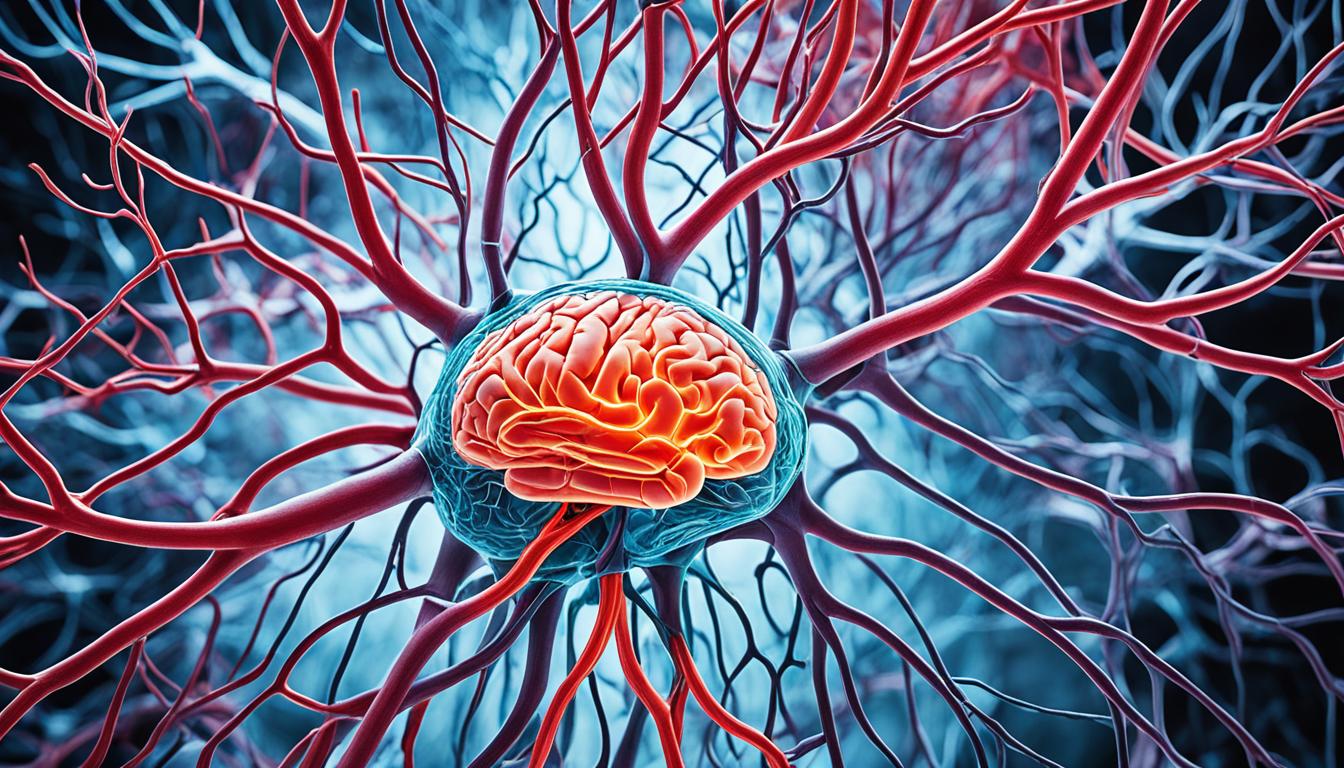
Dural arteriovenous fistulas (dAVFs) are rare and occur in the brain or spinal cord’s tough covering. They cause arteries and veins to connect abnormally. The primary risk group is people aged 50 to 60. These connections can lead to serious conditions like intracranial hemorrhage.
The exact cause of dAVFs isn’t always clear. Yet, one theory is when a venous sinus in the brain gets blocked. This blockage causes blood vessels to form abnormal connections, creating a dAVF.
dAVFs vary in aggressiveness, but they share common symptoms. These include sudden headaches, difficulty walking, seizures, and problems with speech. If you show these symptoms, seek immediate medical help. It might indicate a brain hemorrhage or a seizure.
Imaging tests are crucial for diagnosing dAVFs. CT scans create detailed 3-D images to spot bleeding. MRA makes high-quality images of blood vessels. Cerebral angiography remains the best method to directly see vessel structures.
Treating dAVFs depends on the fistula’s type, where it is, the symptoms’ seriousness, and your health. Treatment may include endovascular therapy, embolization, or surgery. Targeted radiosurgery is also used if other treatments are not an option.
Stem cell therapy is a new area showing potential for treating dAVFs. It’s believed that stem cells can help repair blood vessels and improve blood flow. This offers a promise for a safer, less invasive treatment. But, more research is necessary to confirm its safety and effectiveness.
Key Takeaways:
- Dural arteriovenous fistulas are uncommon and involve abnormal connections between brain or spinal cord blood vessels.
- They can lead to severe symptoms like intracranial hemorrhage and neurological issues.
- The precise cause of dAVFs remains unclear, but a theory involves blockage in the brain’s venous sinus.
- Such conditions may present symptoms such as sudden severe headaches, difficulty walking, and seizures.
- If these symptoms occur, immediate medical evaluation is necessary to rule out serious effects like brain hemorrhage.
- CT scans, MRA, and cerebral angiography are essential for diagnosing dAVFs.
- Treatment options include endovascular therapy, embolization, surgical intervention, or radiosurgery.
- Researchers are exploring stem cell therapy as a novel approach to dAVF treatment.
Diagnosis and Detection of Dural Arteriovenous Fistulas
Finding out someone has dural arteriovenous fistulas (dAVFs) requires special imaging. These images show where they are and how blood flows. Knowing this helps doctors choose the best treatment.
Imaging Techniques for Diagnosis
Computed tomography (CT) scans make detailed 3-D brain pictures. They help spot bleeding in emergencies. This is key for a quick understanding of the patient’s health.
Magnetic resonance angiography (MRA) uses magnets for blood vessel images. It’s great at showing blood flow in the brain. This way, doctors can find the problem areas and see dAVFs.
Cerebral angiography, or arteriogram, is top for dAVF diagnosis. It uses a special dye and X-rays for clear blood vessel views. With this, doctors get a full picture of the dAVF to plan the best treatment.
The Role of Imaging Techniques
CT scans, MR angiography, and cerebral angiography are vital for dAVF diagnosis. They give doctors key details about dAVFs. This information is crucial for picking the right treatment path.
Doctors use these imaging results to create personalized treatment plans. This approach leads to better outcomes and a higher-quality life for the patient.
Ultimately, using advanced imaging to diagnose and detect dural arteriovenous fistulas is critical for proper care.
| Imaging Technique | Advantages | Disadvantages |
|---|---|---|
| Computed Tomography (CT) Scan | Provides 3-D images of the brain Identifies sites of bleeding or hemorrhage |
Limited in evaluating blood flow patterns |
| Magnetic Resonance Angiography (MRA) | Creates detailed images of blood vessels Visualizes blood flow patterns |
May be contraindicated for patients with certain medical devices or conditions |
| Cerebral Angiography | Gold standard for diagnosing dAVFs Provides comprehensive assessment of blood vessels |
Invasive procedure with potential risks |
Treatment Options for Dural Arteriovenous Fistulas
Dural arteriovenous fistulas (dAVFs) have many treatment options. The type and place of the fistula, along with symptoms, age, and health, affect the choice.
Endovascular therapy is one option. It uses coils, glue, or spheres to stop blood flow to the dAVF. This reduces complications from the connection.
Embolization is another choice. It blocks the blood vessels near the dAVF, thus reducing blood flow. It can use materials like coils or liquid agents.
When other methods are not an option, surgical resection might be needed. This involves cutting off or removing the dAVF to stop the abnormal blood flow.
If none of the above work, targeted radiosurgery could be the answer. High-dose radiation is aimed at the dAVF. This gradually closes it off.
The best treatment is decided by a team of experts. Neurosurgeons, radiologists, and neurologists all have a say. They consider the patient’s condition and the pros and cons of each treatment.

